Time slot's time in Taipei (GMT+8)
2025/11/22 11:00-13:30 Room 101 AB
- SYMPOSIUM 1 Tropical Diseases
Neurophysiological Characteristics of Tropical Disease in Asian Oceanian Region
- Time
- Topic
- Speaker
- Moderator
- 11:00-11:30
- Dengue Fever in Taiwan and Asia: the past, current and future
- Speaker:
Yuan-Ting Sun
- Moderator:
Shey-Lin Wu

- Yuan-Ting Sun
- MD PhD
-
Associate Professor, Department of Neurology, National Cheng Kung University Hospital
E-mail:ytsun@ncku.edu.tw
Executive Summary:
Dr. Yuan-Ting Sun, MD, PhD, is an Associate Professor of Neurology at National Cheng Kung University Hospital, Taiwan. She completed her medical and doctoral training at National Cheng Kung University and pursued postdoctoral research at the University of Oxford. With expertise in neuroimmunology, neurogenetics, and neuromuscular disorders, Dr. Sun has made significant contributions to both clinical care and translational research. She currently serves in leadership roles within the Neuromuscular, Neurogenetics, Neuroimmunology, and Rare Disorders Sections of the Taiwan Neurological Society, and her work has been recognized with multiple national and international awards.
Today, Dr. Sun will speak on “Dengue Fever in Taiwan and Asia: the Past, Current and Future.” Her presentation will highlight epidemiological trends and focus on the neurological manifestations of dengue, summarizing past experiences, current challenges, and future strategies for clinical management and prevention. Please join me in welcoming Dr. Yuan-Ting Sun.
Lecture Abstract:
Dengue fever is a rapidly emerging mosquito-borne disease that poses a substantial public health burden in Taiwan and across Asia. While traditionally recognized for its febrile and hemorrhagic manifestations, dengue infection can also involve the nervous system, producing a broad spectrum of neurological complications ranging from encephalitis and myelitis to neuropathies and neuromuscular disorders. These manifestations highlight the complex interplay between host immune responses and viral pathogenesis. In this lecture, I will review the epidemiological trends of dengue in Taiwan and Asia, summarize the neurological complications observed in past and recent outbreaks. I will address future directions for surveillance, clinical management, and prevention strategies, with particular attention to the neurological dimension of this evolving disease.
Dengue fever is a rapidly emerging mosquito-borne disease that poses a substantial public health burden in Taiwan and across Asia. While traditionally recognized for its febrile and hemorrhagic manifestations, dengue infection can also involve the nervous system, producing a broad spectrum of neurological complications ranging from encephalitis and myelitis to neuropathies and neuromuscular disorders. These manifestations highlight the complex interplay between host immune responses and viral pathogenesis. In this lecture, I will review the epidemiological trends of dengue in Taiwan and Asia, summarize the neurological complications observed in past and recent outbreaks. I will address future directions for surveillance, clinical management, and prevention strategies, with particular attention to the neurological dimension of this evolving disease.
- Time
- Topic
- Speaker
- Moderator
- 11:30-12:00
- EMG signals in tropical myositis
- Speaker:
Usha Kant Misra
- Moderator:
Jayantee Kalita
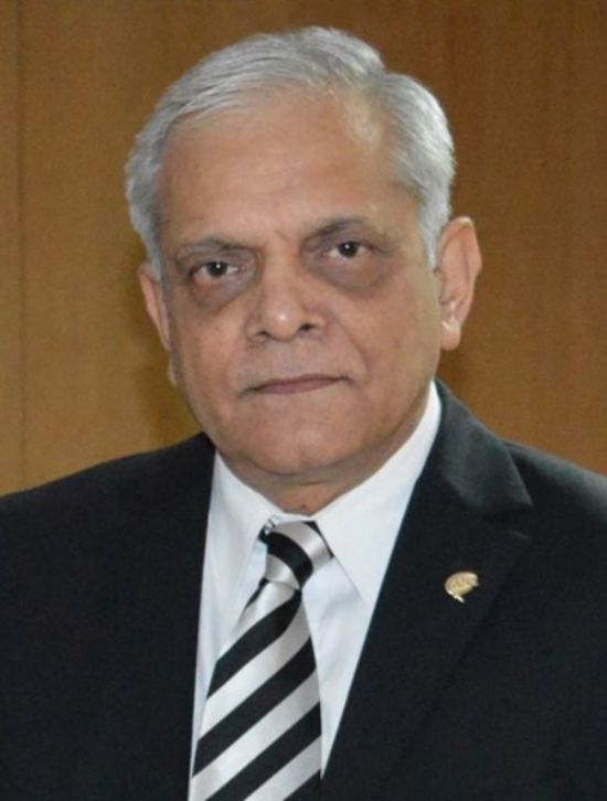
- Usha Kant Misra
- MD, DM, FRCP(Edin, LONDON), FNA, FNASc, FAMS, FAAN
-
T.S. Misra Medical College and Hospital, Lucknow, Professor Emeritus
Apollomedics Super Speciality Hospital, Lucknow, Principal Director, Neurosciences
Vivekananda Polyclinic & Institute of Medical Sciences, Lucknow, Consultant
E-mail:drukmisra@rediffmail.com
Executive Summary:
Prof. U.K. Misra, after graduation in 1973, MD in Medicine in 1978, and DM in Neurology in 1985 from KG Medical College, Lucknow, worked as a Scientist in Neurotoxicology from 1980-1986 and as a Lecturer in Neurology at KGMC in 1986. He trained in Clinical Neurophysiology as a WHO fellow in Sweden, Italy, and the USA. He joined Sanjay Gandhi PGI in 1987 and founded the Department of Neurology, which he headed until 2017. After superannuation, he worked as a Senior Professor of Neurology until 2021. Presently, he is working as Professor Emeritus at TS Misra Medical College, Principal Director at Apollo Medics Super Specialty Hospital, and a Consultant at Vivekanand Polyclinic and Institute of Medical Sciences in Lucknow.
He has made several important and original contributions to CNS infections, including TB meningitis, encephalitis (such as the importance of thalamic lesions in the diagnosis of Japanese encephalitis (JE)), the role of EMG in documenting anterior horn cell involvement in JE, and the spectrum and basis of movement disorders in JE. He has provided valuable information about muscle involvement in dengue and contrasted it with muscle involvement in scrub typhus. In TB meningitis, he has provided information on the role of cerebral salt wasting in the pathogenesis of stroke and the role of aspirin in preventing stroke in TBM.
He has published over 16 clinical trials that have influenced the management of patients and helped in the teaching of students. Dr. Misra has authored 527 papers and 5 books, with citations exceeding 21,387 and an H-index of 74. Based on scientometric analysis of his publications during 1999-2008, he was adjudged the most prolific and impactful author in Neurology and Neurosurgery in India.
Lecture Abstract:
Muscles are affected by a large number of pathogens, including bacteria, viruses, fungi, and parasites, resulting in diverse clinical manifestations. In the post-monsoon period in tropical countries, there are often cases of Acute Flaccid Paralysis of varying severity due to anterior horn cell involvement (Japanese, West Nile encephalitis, enteroviruses), peripheral nerve involvement (GB syndrome triggered by viral illness, e.g., Dengue, enteroviruses), and muscle involvement by Dengue, Scrub Typhus, which have an overlapping clinical picture. The anterior horn cell involvement in JE and WN can be documented by EMG, revealing spontaneous activity in the early phase and neurogenic potentials in the later phase, which normalize in due course. AFP in Dengue is associated with systemic features like thrombocytopenia, liver and kidney dysfunction, coagulopathy, and hypotension. The muscle involvement manifests with myalgia, with or without weakness, and elevated serum creatine kinase (CK). It was considered to be viral myositis, but subsequent studies revealed that the EMG changes were subtle and not diagnostic. Quantitative EMG revealed short duration of MUP, which increased after one month. However, subsequent studies revealed that out of 31 patients, 52% had weakness, and 48% had only serum CK elevation 837 (199-3882) U/L. EMG showed no spontaneous activity and low-amplitude MUP with short duration. Muscle biopsy revealed edema and hemorrhage. In some patients, muscle weakness is due to associated hypokalemia. A similar clinical picture is seen in Scrub Typhus infection, but EMG shows spontaneous activity and myopathic potential, and biopsy showed features of vasculitis. Influenza-related myositis is generally mild, and there is a paucity of reports on EMG findings. Pyogenic infection of muscle (Pyomyositis) results in focal pain and weakness, showing typical changes in imaging and histology. Cysticercoids result in muscle edema and typical findings on imaging. Patients with muscle involvement in infection have diverse manifestations and need a systematic evaluation of clinical, lab data, imaging, EMG, and NCV information.
Muscles are affected by a large number of pathogens, including bacteria, viruses, fungi, and parasites, resulting in diverse clinical manifestations. In the post-monsoon period in tropical countries, there are often cases of Acute Flaccid Paralysis of varying severity due to anterior horn cell involvement (Japanese, West Nile encephalitis, enteroviruses), peripheral nerve involvement (GB syndrome triggered by viral illness, e.g., Dengue, enteroviruses), and muscle involvement by Dengue, Scrub Typhus, which have an overlapping clinical picture. The anterior horn cell involvement in JE and WN can be documented by EMG, revealing spontaneous activity in the early phase and neurogenic potentials in the later phase, which normalize in due course. AFP in Dengue is associated with systemic features like thrombocytopenia, liver and kidney dysfunction, coagulopathy, and hypotension. The muscle involvement manifests with myalgia, with or without weakness, and elevated serum creatine kinase (CK). It was considered to be viral myositis, but subsequent studies revealed that the EMG changes were subtle and not diagnostic. Quantitative EMG revealed short duration of MUP, which increased after one month. However, subsequent studies revealed that out of 31 patients, 52% had weakness, and 48% had only serum CK elevation 837 (199-3882) U/L. EMG showed no spontaneous activity and low-amplitude MUP with short duration. Muscle biopsy revealed edema and hemorrhage. In some patients, muscle weakness is due to associated hypokalemia. A similar clinical picture is seen in Scrub Typhus infection, but EMG shows spontaneous activity and myopathic potential, and biopsy showed features of vasculitis. Influenza-related myositis is generally mild, and there is a paucity of reports on EMG findings. Pyogenic infection of muscle (Pyomyositis) results in focal pain and weakness, showing typical changes in imaging and histology. Cysticercoids result in muscle edema and typical findings on imaging. Patients with muscle involvement in infection have diverse manifestations and need a systematic evaluation of clinical, lab data, imaging, EMG, and NCV information.
- Time
- Topic
- Speaker
- Moderator
- 12:00-12:30
- Characteristic of electrodiagnostic and clinical features of leprosy in Indonesia
- Speaker:
Manfaluty Hakim
- Moderator:
Nghia Tien Trong Hoang

- Manfaluty Hakim
- MD,PhD
-
Head of Clinical Neurophysiology, Neuromuscular Disorder, Epilepsy and Sleep Disorder Division of Neurology Department of Faculty Of Medicine, Universitas Indonesia-Cipto Mengunkusumo Hospital, Jakarta, Department of Neurology, Faculty of Medicine, Univeristas Indonesia
E-mail:hakim_dr@yahoo.com
Executive Summary:
Manfaluthy Hakim, neurologist from neurology department of Universitas Indonesia, Jakarta, Indonesia. Currently, work as Head of Division of Clinical Neurophysiology, Neuromuscular Disorder, Epilepsy and Sleep Disorder, Neurology Department Faculty of Medicine, Universitas Indonesia and National General Hospital Dr. Cipto Mangunkusumo.
Manfaluthy Hakim, neurologist from neurology department of Universitas Indonesia, Jakarta, Indonesia. Currently, work as Head of Division of Clinical Neurophysiology, Neuromuscular Disorder, Epilepsy and Sleep Disorder, Neurology Department Faculty of Medicine, Universitas Indonesia and National General Hospital Dr. Cipto Mangunkusumo.
Lecture Abstract:
Electrodiagnostic and Clinical Features of Leprosy in Indonesia
Manfaluthy Hakim
Abstract
Leprosy is a common, treatable cause of peripheral neuropathy in many tropical and subtropical countries. Mycobacterium leprae has a distinct predilection for areas of low body temperature and a unique tropism for nerves. Leprosy can lead to skin lesions and peripheral nerve damage in the form of neuropathy if untreated. It frequently afflicts the ulnar nerve possibly due to its superficial location. The frequent delays in the diagnosis of leprosy allows the progression of nerve damage, often resulting in deformity. It primarily targets the skin and peripheral nerves, but in some cases, depending on the spectrum of the disease (lepromatous) and delay in diagnosis, leprosy can affect mucosa of the upper respiratory tract and the eyes.
Based on WHO data from 2019, Indonesia is in the top three countries with the most significant contributors to leprosy cases after India and Brazil. World Health Organization (WHO) data, the number of leprosy patients worldwide in 2019 reached 202,226. Out of this total, 143,787 cases were reported in Southeast Asia.
The diagnosis of leprosy base on clinical examination, serology, biopsy and neurophysiological studies. The nerve injury is difficult to assess by conventional neurophysiological techniques such as electroneuromyography, although late latency evaluations such as H-reflex, blink reflex, the F-wave and sympathetic skin response are useful in their detection. Neurophysiological studies in leprosy reveal asymmetrical nerve involvement, often manifesting as mononeuropathy or mononeuritis multiplex, especially affecting sensory nerves.


