Time slot's time in Taipei (GMT+8)
2025/11/22 14:00-17:30 Room 101 AB
- SYMPOSIUM 3&7 Neuromuscular Disorders
Expanding Frontier of Clinical Neurophysiology in Diagnosis and Treatment of Neuromuscular Diseases
- Time
- Topic
- Speaker
- Moderator
- 14:00-14:30
- Advances in neurophysiological biomarkers for disease diagnosis and progression of ALS
- Speaker:
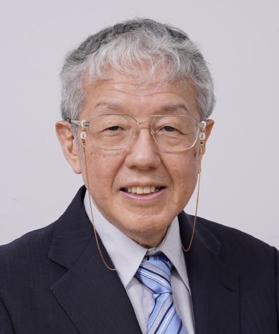
Masahiro Sonoo
- Moderator:
Yi-Chung Lee
- Masahiro Sonoo
- MD
-
Professor Emeritus, Department of Neurology, Teikyo University School of Medicine
Professor, Department of Orthoptics, Faculty of Medical Technology, Teikyo University
E-mail:sonoom@med.teikyo-u.ac.jp
Executive Summary:
Dr. Masahiro Sonoo graduated from University of Tokyo in 1982, and started his neurology training at Tokyo University Hospital in Tokyo, Japan. He studied at Prof. Erik Stålberg’s laboratory in Uppsala, Sweden for 1 year from 1991. Since he came back to Japan, he has worked for department of Neurology, Teikyo University School of medicine, Tokyo, Japan for more than 30 years, and was Professor and Chairman of the department from 2011 to 2023. Since 2023, he has been Professor Emeritus of Teikyo University, as well as Professor of Faculty of Medical Technology and Affiliate Professor of Department of Neurology, Teikyo University. He is an auditor and former director of Japanese Society of Neurology, and also is an auditor and former director of Japanese Society of Clinical Neurophysiology. He is an editorial board member of Muscle and Nerve, Clinical Neurophysiology Practice, and Clinical Neurophysiology.
His research interests include concentric needle EMG such as invention of Size Index in quantitative EMG, quantitative surface EMG named Clustering Index Method, single-fiber EMG where he established normal values of concentric SFEMG by a multi-center study, SEPs where he determined generators of several components, nerve conduction studies including far-field potentials in CMAP, determination of myotomes, new selectivity in ALS such as split finger and arm sparing/weak shoulder, functional neurological disorders where he discovered a number of new positive signs of functional weakness including abductor test, wrist flexor test, and gluteus maximus test, etc.
Lecture Abstract:
Neurophysiological biomarkers for Amyotrophic lateral sclerosis (ALS) would be classified into those for lower motor neurons (LMNs) and for upper motor neurons (UMNs). I do not major in UMN biomarkers, which probably remain within research interest for the present. Therefore, I mainly talk about LMN biomarkers in this lecture. LMN involvements in ALS can be evaluated either by clinical signs or by electrophysiological examinations. Among clinical signs, selective muscle involvements, i.e. split phenomena, draw the attention of recent researchers. These include split hand, split hand plus, split leg, split elbow, split finger, and arm sparing/weak shoulder, some of which are not supported by following studies. Split hand has been also evaluated neurophysiologically as split hand index. Conventionally, LMN involvements in ALS has been evaluated by needle EMG. Widespread denervation together with changes of voluntary activities is a gold standard for ALS diagnosis, although it may not be evident in the early stage. Spontaneous activities in the trapezius muscle are highly specific for ALS at least in comparison with cervical spondylosis. Decremental response in repetitive nerve stimulation is frequently observed in ALS. This is usually believed to reflect immature sprouts in ongoing reinnervation, but we suggested that this is not the sole mechanism. Decremental response in the trapezius muscle is specific for ALS in comparison with cervical spondylotic amyotrophy. The importance of surrogate markers for disease progression is increasing in this era of emerging treatments. Needle EMG is not suited for this purpose because of its invasiveness. Compound muscle action potential (CMAP) amplitude would be the simplest marker, although low reproducibility would be its limitation. Far-field potential of the CMAP can overcome this drawback. Motor unit number estimate (MUNE) and MUNIX can evaluate the number of remaining motor neurons.
Neurophysiological biomarkers for Amyotrophic lateral sclerosis (ALS) would be classified into those for lower motor neurons (LMNs) and for upper motor neurons (UMNs). I do not major in UMN biomarkers, which probably remain within research interest for the present. Therefore, I mainly talk about LMN biomarkers in this lecture. LMN involvements in ALS can be evaluated either by clinical signs or by electrophysiological examinations. Among clinical signs, selective muscle involvements, i.e. split phenomena, draw the attention of recent researchers. These include split hand, split hand plus, split leg, split elbow, split finger, and arm sparing/weak shoulder, some of which are not supported by following studies. Split hand has been also evaluated neurophysiologically as split hand index. Conventionally, LMN involvements in ALS has been evaluated by needle EMG. Widespread denervation together with changes of voluntary activities is a gold standard for ALS diagnosis, although it may not be evident in the early stage. Spontaneous activities in the trapezius muscle are highly specific for ALS at least in comparison with cervical spondylosis. Decremental response in repetitive nerve stimulation is frequently observed in ALS. This is usually believed to reflect immature sprouts in ongoing reinnervation, but we suggested that this is not the sole mechanism. Decremental response in the trapezius muscle is specific for ALS in comparison with cervical spondylotic amyotrophy. The importance of surrogate markers for disease progression is increasing in this era of emerging treatments. Needle EMG is not suited for this purpose because of its invasiveness. Compound muscle action potential (CMAP) amplitude would be the simplest marker, although low reproducibility would be its limitation. Far-field potential of the CMAP can overcome this drawback. Motor unit number estimate (MUNE) and MUNIX can evaluate the number of remaining motor neurons.
- Time
- Topic
- Speaker
- Moderator
- 14:30-15:00
- Emerging neuroimaging techniques for the structural and functional changes of ALS
- Speaker:
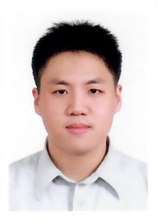
Sung-Ju Hsueh
- Moderator:
Chih-Chao Yang
- Sung-Ju Hsueh
- MD, MSc
-
Attending Physician, Department of Neurology, National Taiwan University Hospital Bei-Hu Branch, Taiwan
Adjunct Lecturer, Department of Neurology, National Taiwan University College of Medicine, Taiwan
E-mail:b95401008@ntu.edu.tw
Executive Summary:
Dr. Sung-Ju Hsueh received his MD degree from the School of Medicine, National Taiwan University, Taiwan. He received clinical neurology specialist training at National Taiwan University Hospital (NTUH), and is a board-certified neurologist in Taiwan. He also received an MSc degree from the Institute of Clinical Medicine, National Taiwan University, for his study on the imaging markers of ALS.
Currently, Dr. Hsueh works at the NTUH Bei-Hu branch as an attending neurologist. He is also an adjunct Instructor at the Department of Neurology, College of Medicine, National Taiwan University. His research interest includes motor neuron disease, peripheral neuropathy, neuromuscular disease, and electrophysiology.
Lecture Abstract:
Amyotrophic lateral sclerosis (ALS) is a neurodegenerative disease with prominent signs of neurodegeneration in the central nervous system (CNS). Many imaging techniques had been utilized to study the pathophysiological process of ALS, revealing widespread changes in the CNS. Furthermore, imaging studies had also identified structural and functional changes contributing to the non-motor symptoms of ALS, such as cognitive impairment. In patients with genetic ALS and carriers, neuroimaging studies had identified sequential changes in the CNS and could serve as predictive markers for phenoconversion.
Recently, advanced imaging approaches, such as metabolic, molecular, inflammatory, and glymphatic imaging, had identified novel pathological pathways and features contributing to the development of ALS. In this session, I will discuss the recent developments and emerging neuroimaging techniques for the study of ALS.
- Time
- Topic
- Speaker
- Moderator
- 15:00-15:30
- Nerve Excitability: A Multidisciplinary Initiative Translatio
- Speaker:
Cindy Shin-Yi Lin
- Moderator:
Jia-Ying Sung
- Cindy Shin-Yi Lin
- PhD, MESci(Biomedical), BE
-
The Kam Ling Barbara Lo Chair in Neurodegenerative Disorders, Central Clinical School, Faculty of Medicine and Health, Brain and Mind Centre, University of Sydney
E-mail:cindy.lin@sydney.edu.au
Executive Summary:
Professor Cindy (Shin-Yi) Lin is a leading international expert in neurophysiology and neuromuscular disorders, currently serving as the inaugural Kam Ling Barbara Lo Chair in Neurodegenerative Disorders at the University of Sydney’s Brain & Mind Centre. With postdoctoral training at University College London’s Institute of Neurology and an outstanding track record in both research and education, Professor Lin has pioneered the clinical application and translation of nerve excitability techniques in the diagnosis and management of peripheral neuropathies.
She has authored over 150 peer-reviewed publications and book chapters with current H-index of 50, including foundational texts and international guidelines in clinical neurophysiology. Professor Lin’s research encompasses early detection and detailed phenotyping of hereditary, metabolic, and acquired neuropathies, as well as neurophysiological markers of disease progression and therapeutic response. Her multidisciplinary and highly collaborative approach bridges neurophysiology, clinical neurology, and computational modeling, with global research partnerships in the UK, Japan, France, Switzerland, and Taiwan.
Professor Lin has delivered invited lectures and workshops worldwide, training neurologists in nerve excitability methodology. Her leadership in research has been recognized by numerous prestigious grants and awards, including the NHMRC CJ Martin Fellowship and CSL Behring Interlaken Leadership Award. She serves on editorial boards for leading journals such as the Journal of Neurology, Neurosurgery and Psychiatry and Frontiers in Neurology. Professor Lin’s mission is to be a true translational scientist and advocate for innovation in clinical neurophysiology.
Lecture Abstract:
Nerve excitability techniques (NET) have emerged as advanced tools for the assessment and diagnosis of peripheral neuropathies, providing insights beyond traditional nerve conduction studies (NCS). By applying controlled electrical stimuli and monitoring the nerve's response, these techniques evaluate axonal membrane properties, including voltage-gated ion channel function, membrane polarization, and the activity of energy-dependent ion pumps. Unlike conventional methods, NET offers a dynamic profile of axonal health by measuring parameters such as threshold electrotonus, strength–duration time constant, rheobase, and recovery cycles. This approach enables the detection of functional changes in ion channel activity and nodal/internodal properties, which are essential for understanding the pathophysiology of various peripheral nerve disorders.
Clinically, NET facilitates early diagnosis and phenotyping of neuropathies, including hereditary, metabolic, and acquired disorders. The method can reveal discrete alterations in nerve properties before the onset of overt symptoms or conduction block, supporting timely intervention. Additionally, it provides a sensitive marker for monitoring disease progression and response to therapy, as well as investigating peripheral manifestations of central nervous system channelopathies.
The translation of NET into multidisciplinary clinical practice underscores its value as a non-invasive, quantitative, and reproducible diagnostic tool. By integrating neurophysiology, clinical neurology, and computational modeling, nerve excitability studies enhance our ability to unravel complex disease mechanisms and personalize patient care. Ongoing research is refining these protocols and further establishing their place in the diagnostic algorithm for peripheral neuropathies, broadening NET application from research laboratories to routine clinical neurology.
- Time
- Topic
- Speaker
- Moderator
- 16:00-16:30
- Pathological basis of nerve conduction abnormalities and its application to the diagnosis of peripheral neuropathy
- Speaker:
Koike Haruki
- Moderator:
Yuh-Cherng Guo
- Koike Haruki
- MD, PhD
-
Professor, Department of Neurology, Saga University
E-mail:haruki.koike@gmail.com
Executive Summary:
Professor Haruki Koike received his medical degree from the Tohoku University and completed postgraduate doctoral course in the Department of Neurology at Nagoya University Graduate School of Medicine. His clinical and research interests include the clinicopathological and therapeutic aspects of various kinds of inflammatory and non-inflammatory neuropathies, including chronic inflammatory demyelinating polyneuropathy, Guillain-Barré syndrome, nutritional neuropathies, hereditary transthyretin (ATTRv) amyloidosis, and vasculitic neuropathies. He is a director of Japanese Peripheral Nerve Society, Japanese Society of Neuropathology, and Japanese Society of Amyloidosis.
Professor Haruki Koike received his medical degree from the Tohoku University and completed postgraduate doctoral course in the Department of Neurology at Nagoya University Graduate School of Medicine. His clinical and research interests include the clinicopathological and therapeutic aspects of various kinds of inflammatory and non-inflammatory neuropathies, including chronic inflammatory demyelinating polyneuropathy, Guillain-Barré syndrome, nutritional neuropathies, hereditary transthyretin (ATTRv) amyloidosis, and vasculitic neuropathies. He is a director of Japanese Peripheral Nerve Society, Japanese Society of Neuropathology, and Japanese Society of Amyloidosis.
Lecture Abstract:
Peripheral neuropathy is caused by a variety of diseases, including diabetes mellitus, alcoholism, nutritional deficiency, and autoimmune diseases represented by Guillain-Barré syndrome (GBS) and chronic inflammatory demyelinating polyneuropathy (CIDP). Based on the characteristics of electrophysiological findings, GBS is mainly categorized into a demyelinating subtype known as acute inflammatory demyelinating polyneuropathy (AIDP) and an axonal subtype known as acute motor axonal neuropathy (AMAN). Early reports of pathological findings in patients with AIDP and CIDP identified macrophages as important players in the pathogenesis of neuropathy because demyelination resulting from the phagocytosis of myelin by macrophages was considered as the cause of aberrant nerve conduction in these diseases. Later studies have focused on the search for autoantibodies directed against peripheral nerve components for clarifying the mechanisms of nerve conduction abnormalities in patients with immune-mediated neuropathies. For example, a concept that molecular mimicry of gangliosides in the peripheral nervous system to surface epitopes of exogenous pathogens results in the production of autoantibodies has been established in patients with AMAN. In contrast, the mechanisms of neuropathy in cases with classical macrophage-induced demyelination are still to be elucidated. In addition, recent studies have suggested the association of IgG4 autoantibodies directed against paranodal junction proteins, such as neurofascin 155, contactin 1, and Caspr1, to some of the patients previously diagnosed as CIDP. Although patients with these antibodies meet the electrodiagnostic criteria for CIDP, classical macrophage-induced demyelination is not observed in these patients. Paranodal axo-glial dissection resulting from attachment of IgG4 antibodies to paranodal junctions plays a pivotal role in the mechanisms of neuropathy. These findings led to the emergence of the concept of autoimmune nodopathy, which is distinct from CIDP.
Peripheral neuropathy is caused by a variety of diseases, including diabetes mellitus, alcoholism, nutritional deficiency, and autoimmune diseases represented by Guillain-Barré syndrome (GBS) and chronic inflammatory demyelinating polyneuropathy (CIDP). Based on the characteristics of electrophysiological findings, GBS is mainly categorized into a demyelinating subtype known as acute inflammatory demyelinating polyneuropathy (AIDP) and an axonal subtype known as acute motor axonal neuropathy (AMAN). Early reports of pathological findings in patients with AIDP and CIDP identified macrophages as important players in the pathogenesis of neuropathy because demyelination resulting from the phagocytosis of myelin by macrophages was considered as the cause of aberrant nerve conduction in these diseases. Later studies have focused on the search for autoantibodies directed against peripheral nerve components for clarifying the mechanisms of nerve conduction abnormalities in patients with immune-mediated neuropathies. For example, a concept that molecular mimicry of gangliosides in the peripheral nervous system to surface epitopes of exogenous pathogens results in the production of autoantibodies has been established in patients with AMAN. In contrast, the mechanisms of neuropathy in cases with classical macrophage-induced demyelination are still to be elucidated. In addition, recent studies have suggested the association of IgG4 autoantibodies directed against paranodal junction proteins, such as neurofascin 155, contactin 1, and Caspr1, to some of the patients previously diagnosed as CIDP. Although patients with these antibodies meet the electrodiagnostic criteria for CIDP, classical macrophage-induced demyelination is not observed in these patients. Paranodal axo-glial dissection resulting from attachment of IgG4 antibodies to paranodal junctions plays a pivotal role in the mechanisms of neuropathy. These findings led to the emergence of the concept of autoimmune nodopathy, which is distinct from CIDP.
- Time
- Topic
- Speaker
- Moderator
- 16:30-17:00
- Clinical and neurophysiological evaluations of myotonic disorder
- Speaker:
Yuh-Cherng Guo
- Moderator:
Kuan-Lin Lai
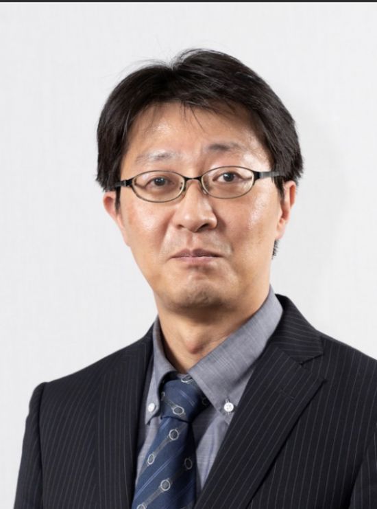
- Yuh-Cherng Guo
- MSc
-
Director of Section of Neuroimmune and Neurogenetic disorders, Department of Neurology, China Medical University Hospital, China Medical University Hospital
E-mail:cmtpmp22@gmail.com
Executive Summary:
Dr. Guo was graduated from Kaohsiung Medical University, Taiwan and completed master’s degree from Graduate Institute of Medicine, College of Medicine, Kaohsiung Medical University. He has finished the training of resident at Department of Neurology, Neurological Institute, Taipei Veterans General Hospital. Then he moved to Kaohsiung Medical University Hospital as an attending physician. Now he is an attending physician and the director of Section of Neuroimmune and Neurogenetic Disorders at Department of Neurology, China Medical University Hospital, Taichung.
Dr. Guo’s interests focus on the field of neuroimmunology, neurophysiology of neuropathies, neuromuscular junction disorders and myopathies, and genetic study of neurological neuromuscular diseases. He has been working at the diagnosis and management of multiple sclerosis, neuromyelitis optica spectrum disorder (NMOSD), autoimmune encephalitis, dysimmune neuropathies, myasthenia gravis and inherited neuropathies and myopathies.
Research Interests
Neuroimmunology, Neurophysiology, Pathology of neuropathies and myopathies
Lecture Abstract:
Myotonic disorders are characterized by the failure of muscles relax after contraction, or myotonia. The genetic defects involve the muscular isoforms of the ion channels. These diseases are inherited myopathies divided into dystrophic myotonias and non-dystrophic myotonias. The most common dystrophic myopathy is myotonic dystrophy type I (DM1), are multisystemic disorders that include progressive muscle weakness and wasting, in addition to myotonia.
Non-dystrophic myotonias (NDM) do not have significant progressive muscle wasting or multisystemic complications. It is a group of autosomal dominant or recessive diseases caused by mutations in genes encoding ion channels (chloride or sodium channels) that participate in the generation and control of the skeletal muscle action potentials. The symptoms of NDM start in early to late childhood or the teenage years including episodes of myotonias, stiffness, cramps, locking ot tightness. Myotonia can happen in the legs, arms, hands, abdomen, back and face (including the tongue). Myotonias can be triggered by temperature, movement after rest or with exercise.
In myotonia congenita, myotonia tends to affect the leg muscles mostly, but can happen in other muscles too. Myotonia can be improved by activating the muscles repeatedly, a process known as the “warm-up phenomenon”.
In paramyotonia congenita, myotonia involves the hands and face muscles the most, but can also happen in the legs and other muscles. Unlike myotonia congenita, myotonia gets worse when repeatedly using the muscles. Som patients with paramyotonia congenita can also experience episodes of muscle weakness or paralysis which might last from a few minutes to hours.
There are two main medications used to treat myotonia: mexiletine and lamotrigine. Mexiletine can be taken regularly or when needed to manage muscle stiffness. An electrocardiogram (ECG) is needed before the use of mexiletine.
- Time
- Topic
- Speaker
- Moderator
- 17:00-17:30
- Neurophysiological techniques for localizing brachial and lumbosacral plexopathy
- Speaker:
Jorge Eduardo Gutiérrez
- Moderator:
Chien-Hsu Lai
- Jorge Eduardo Gutiérrez
- MD, MSc
-
Medical director, Potenciales Center of Electrodiagnosis
E-mail:jegutier@gmail.com
Executive Summary:
Dr. Jorge Gutierrez completed his specialist training in Physical Medicine and Rehabilitation at Universidad del Valle in Cali, Colombia. He established the EMG laboratory at Universidad del Cauca in Popayán, Colombia, and founded the PM&R service at the same institution. Later, Dr. Gutierrez led the EMG laboratory at Hospital Universitario del Valle in Cali.
He held the position of President of the Colombian Society of PM&R and served as President of the Colombian Association of Electrodiagnostic Medicine. Additionally, he acted as Treasurer and subsequently President of the Latin-American chapter of the International Federation of Clinical Neurophysiology (IFCN). Dr. Gutierrez has also been an ExCo member of the International Society of Physical and Rehabilitation Medicine (ISPRM) and served as President of the ISPRM 2023 World Congress.
Dr. Gutierrez is the current secretary of the IFCN ExCo and co-chair of the scientific program committee for the 2026 International Congress of Clinical Neurophysiology in Cartagena, Colombia.
He specializes in electrophysiology and the pathophysiology of peripheral nerve disorders, particularly those caused by trauma. He began his work in intraoperative monitoring in the early 1990s and established himself as a leading pioneer in this field within his country. Other areas of focus include evaluation of plexopathies, use of somatosensory evoked potentials for diagnosing sensory mononeuropathies, neurophysiological assessments of the pelvic floor, and neuromuscular disorders linked to cancer and its treatments.
DR. Gutierrez has authored several publications and book chapters on Electrodiagnosis and is regularly invited to participate in international conferences focused on clinical neurophysiology.
Lecture Abstract:
Plexopathies, defined as dysfunctions of the brachial or lumbosacral plexus, are complex neurological disorders that can result from trauma, neoplasms, radiation, inflammation, or compressive etiologies. Accurate diagnosis is often challenging due to the anatomical intricacy of the plexuses and the overlapping clinical presentations with radiculopathies and peripheral neuropathies. Electrodiagnostic studies, including nerve conduction studies (NCS) and electromyography (EMG), serve as indispensable tools in the evaluation of plexopathies, offering both localization and characterization of the lesion.
NCS are instrumental in differentiating axonal from demyelinating processes, identifying conduction blocks, and assessing the extent of nerve involvement. They can reveal asymmetrical or patchy abnormalities that are characteristic of plexopathies, as opposed to the more uniform changes seen in peripheral neuropathies. EMG complements NCS by detecting denervation in limb muscles supplied by the affected plexus and can help distinguish between pre- and post-ganglionic lesions. The pattern of abnormal spontaneous activity and the distribution of affected muscles on EMG provide clues to the site and severity of plexus involvement.
Electrodiagnostic studies also play a crucial role in prognostication and treatment planning. Serial studies can monitor disease progression or recovery, informing rehabilitation strategies. Furthermore, these modalities may identify occult injuries not evident on imaging and can be particularly valuable when MRI or ultrasound is inconclusive.
In summary, electrodiagnostic studies are vital in the assessment of plexopathies, aiding in precise localization, etiological differentiation, and management decisions. Their integration with clinical and imaging findings ensures a comprehensive diagnostic approach, ultimately improving patient outcomes in this diverse and challenging group of disorders.


