Time slot's time in Taipei (GMT+8)
2025/11/22 14:00-17:30 Room 101 CD
- SYMPOSIUM 4&8 Epilepsy
Update the Clinical Application of EEG and MEG in Epilepsy
- Time
- Topic
- Speaker
- Moderator
- 14:00-14:30
- Non-Convulsive Status Epilepticus: revisited EEG criteria
- Speaker:
Tan Hui Jan
- Moderator:
Rabani Remli

- Tan Hui Jan
- FRCP
-
Head of Neurology Unit and Consultant Neurologist, Faculty of Medicine, National University of Malaysia
E-mail:tanhuijan@gmail.com
Executive Summary:
Dr Tan Hui Jan is a Consultant Neurologist and Head of the Neurology Unit at the National University of Malaysia (UKM). After obtaining her Membership from the Royal College of Physicians (UK), she trained at St Vincent’s Hospital, Melbourne. Dr Tan is actively involved in clinical service, teaching, and research, with a focus on epilepsy, electroencephalogram and headache disorders. She has led and collaborated on numerous research projects, published extensively in peer-reviewed journals, and played a key role in curriculum development and postgraduate training. She has published extensively in peer-reviewed journals and supervises postgraduate students across multiple disciplines. She has contributed to national and international neurology forums and societies. She was awarded for Teaching and Innovation for developing a mobile app to support epilepsy patients. Dr Tan continues in her efforts to improve neurological care, education, and patient outcomes.
Dr Tan Hui Jan is a Consultant Neurologist and Head of the Neurology Unit at the National University of Malaysia (UKM). After obtaining her Membership from the Royal College of Physicians (UK), she trained at St Vincent’s Hospital, Melbourne. Dr Tan is actively involved in clinical service, teaching, and research, with a focus on epilepsy, electroencephalogram and headache disorders. She has led and collaborated on numerous research projects, published extensively in peer-reviewed journals, and played a key role in curriculum development and postgraduate training. She has published extensively in peer-reviewed journals and supervises postgraduate students across multiple disciplines. She has contributed to national and international neurology forums and societies. She was awarded for Teaching and Innovation for developing a mobile app to support epilepsy patients. Dr Tan continues in her efforts to improve neurological care, education, and patient outcomes.
Lecture Abstract:
Non-convulsive status epilepticus (NCSE) is a clinically and electrographically defined condition characterized by prolonged epileptic activity without prominent motor symptoms. It represents a significant portion of status epilepticus (SE) cases, particularly in the elderly, and is associated with diagnostic challenges. Electroencephalography (EEG) plays a pivotal role in the detection and confirmation of NCSE. The Salzburg EEG criteria provide a structured and validated approach for diagnosing NCSE. The modified Salzburg Consensus Criteria have been updated to enhance diagnostic sensitivity, particularly in critically ill patients or those with impaired consciousness. Nevertheless, mimickers such as metabolic encephalopathies and post-hypoxic states require careful differentiation. This presentation reviews the epidemiology, clinical spectrum, and key EEG findings in NCSE, and practical applications in both emergent and continuous EEG monitoring settings. Recognizing NCSE promptly is essential for timely intervention and improved neurological outcomes.
Non-convulsive status epilepticus (NCSE) is a clinically and electrographically defined condition characterized by prolonged epileptic activity without prominent motor symptoms. It represents a significant portion of status epilepticus (SE) cases, particularly in the elderly, and is associated with diagnostic challenges. Electroencephalography (EEG) plays a pivotal role in the detection and confirmation of NCSE. The Salzburg EEG criteria provide a structured and validated approach for diagnosing NCSE. The modified Salzburg Consensus Criteria have been updated to enhance diagnostic sensitivity, particularly in critically ill patients or those with impaired consciousness. Nevertheless, mimickers such as metabolic encephalopathies and post-hypoxic states require careful differentiation. This presentation reviews the epidemiology, clinical spectrum, and key EEG findings in NCSE, and practical applications in both emergent and continuous EEG monitoring settings. Recognizing NCSE promptly is essential for timely intervention and improved neurological outcomes.
- Time
- Topic
- Speaker
- Moderator
- 14:30-15:00
- Electric Source Imaging: the principles and clinical application
- Speaker:
Margitta Seeck
- Moderator:
Fa-Hsuan Lin

- Margitta Seeck
- MD
-
Director of EEG and Epilepsy Unit, University Hospital of Geneva
E-mail:margitta.seeck@hug.ch
Executive Summary:
Dr Margitta Seeck is full professor of Neurology at the University of Geneva, Switzerland. She is director of the presurgical epilepsy program Geneva-Lausanne, which became a reference center for difficult-to-treat epilepsies in adults and children. She also heads the in- and outpatient unit of the EEG and Epilepsy Unit of the University Hospital of Geneva and established the first "first seizure clinic" with inpatient monitoring in the world. She is past president of the Swiss Neurophysiological Society but she is still actively involved in teaching of EEG and epileptology. As Editor-in-chief for Clinical Neurophysiology Practice she is also part of the Executive Committee of the International Federation of Neurology. Her main interests are epilepsy surgery, EEG and EEG-based imaging in epilepsy, neurophysiology of intracranial EEG recordings. She is author of more than 300 papers and bookchapters on epilepsy and EEG in national and international journals, serves on the board of the European Academy of Neurology and several editorial boards of epilepsy journals, and is expert for the Swiss national science foundation and research agencies of other European and non-European countries.
Dr Margitta Seeck is full professor of Neurology at the University of Geneva, Switzerland. She is director of the presurgical epilepsy program Geneva-Lausanne, which became a reference center for difficult-to-treat epilepsies in adults and children. She also heads the in- and outpatient unit of the EEG and Epilepsy Unit of the University Hospital of Geneva and established the first "first seizure clinic" with inpatient monitoring in the world. She is past president of the Swiss Neurophysiological Society but she is still actively involved in teaching of EEG and epileptology. As Editor-in-chief for Clinical Neurophysiology Practice she is also part of the Executive Committee of the International Federation of Neurology. Her main interests are epilepsy surgery, EEG and EEG-based imaging in epilepsy, neurophysiology of intracranial EEG recordings. She is author of more than 300 papers and bookchapters on epilepsy and EEG in national and international journals, serves on the board of the European Academy of Neurology and several editorial boards of epilepsy journals, and is expert for the Swiss national science foundation and research agencies of other European and non-European countries.
Lecture Abstract:
Electrical source imaging (ESI) is an elegant method, in the hands of neurologists, to identify the source of the underlying signal. It is mainly utilized for presurgical epilepsy evaluation, and several reviews have shown that the precision is high, even on a sublobar level. However, ESI can be also used for the localization of vital functions. I will provide an overview of the latest developments, advances and pitfalls.
Electrical source imaging (ESI) is an elegant method, in the hands of neurologists, to identify the source of the underlying signal. It is mainly utilized for presurgical epilepsy evaluation, and several reviews have shown that the precision is high, even on a sublobar level. However, ESI can be also used for the localization of vital functions. I will provide an overview of the latest developments, advances and pitfalls.
- Time
- Topic
- Speaker
- Moderator
- 15:00-15:30
- The intracranial EEG source localization
- Speaker:
Fa-Hsuan Lin
- Moderator:
Yo-Tsen Liu
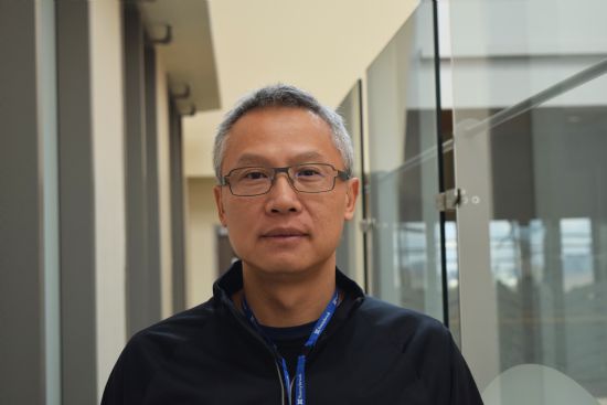
- Fa-Hsuan Lin
- PhD
-
Senior scientist, Sunnybrook Research Institute
Professor, Department of Medical Biophysics, University of Toronto
E-mail:fhlin@sri.utoronto.ca
Executive Summary:
Dr. Fa-Hsuan Lin is a senior scientist at Sunnybrook Research Institute and a professor in the Department of Medical Biophysics at the University of Toronto. Dr. Lin's research focuses on human neuroimaging methodological development, including magnetic resonance imaging, electroencephalography, and magnetoencephalography. The current research focus of Dr. Lin's lab is integrating neuroimaging and neuromodulation, including transcranial magnetic stimulation and complex naturalistic audiovisual stimuli, to establish a closed-loop platform to monitor spatiotemporal brain activity and guide its trajectory toward a healthy one.
Dr. Fa-Hsuan Lin is a senior scientist at Sunnybrook Research Institute and a professor in the Department of Medical Biophysics at the University of Toronto. Dr. Lin's research focuses on human neuroimaging methodological development, including magnetic resonance imaging, electroencephalography, and magnetoencephalography. The current research focus of Dr. Lin's lab is integrating neuroimaging and neuromodulation, including transcranial magnetic stimulation and complex naturalistic audiovisual stimuli, to establish a closed-loop platform to monitor spatiotemporal brain activity and guide its trajectory toward a healthy one.
Lecture Abstract:
Intracranial stereoelectroencephalography (sEEG) provides unsurpassed sensitivity and specificity for human neurophysiology. However, functional mapping of brain functions has been limited because the implantations have sparse coverage and differ greatly across individuals. Here, we present a distributed, anatomically realistic SEEG source modeling approach for within- and between-subject analyses. In the first demonstration of this method, we estimated the spatial distribution of intracranial event-related potentials and high broadband gamma activity (HBBG), a putative correlate of local neural firing. Source modeling suggests that the sensitivity and signal-to-noise ratio were linearly inversely related to the minimal distance between the brain location and electrode contacts (slope ≈−3.6). The signal-to-noise ratio and sensitivity in the thalamus and brain stem were comparable to those locations at the vicinity of electrode contact implantation. The HGGB source estimates were remarkably consistent with analyses of intracranial-contact data.
We then translated the SEEG source modeling to epilepsy recordings to mitigate the challenge of the sampling bias, where no SEEG recording is taken within a circumscribed epileptogenic zone (EZ). Using a retrospective leave-one-out data sub-sampling, we evaluated the sensitivity and specificity of the current estimates using MRI after surgical resection or radio-frequency ablation. The sensitivity and specificity in detecting the EZ were indistinguishable from either the data from all electrodes or the sub-sampled data (rank sum test: rank sum = 23719, p = 0.13) when at least one remaining electrode contact was no more than 20 mm away.
Our results in neuroscience study and clinical application show that the distributed sEEG source modeling is a powerful neuroimaging tool to enable anatomically-normalized functional mapping of the human brain. Clinically, the distributed neuronal current estimates of ictal SEEG data can mitigate the challenge of delineating the boundary of the EZ in cases of missing an electrode implanted within the EZ and a required second SEEG exploration. This modeling tool can assist clinicians in inferring the EZ by allowing for more flexible planning of the electrode implantation route and minimizing the number of electrodes.
- Time
- Topic
- Speaker
- Moderator
- 16:00-16:30
- MEG technology advancements and clinical applications
- Speaker:
Nobukazu Nakasato
- Moderator:
Yung-Yang Lin
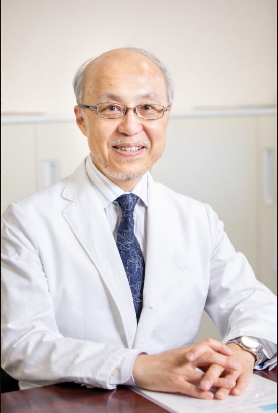
- Nobukazu Nakasato
- MD, PhD
-
Professor, Tohoku University Graduate School of Medicine
Director, Kohan Hospital
E-mail:nkst@tohoku.ac.jp
Executive Summary:
Dr. Nobukazu Nakasato graduated from Tohoku University School of Medicine in 1984. In parallel with neurosurgical residency program, he has also begun research of magnetoencephalography (MEG). Dr. Nakasato studied clinical application of MEG as a part of comprehensive epilepsy program in UCLA from 1989 to 1991. In 1993, Dr. Nakasato introduced the first helmet-shaped MEG system in Japan. In 2010, he became a professor at Tohoku University and founded the Department of Epilepsy. In retired from Tohoku University in 2025 and received the title of Emeritus Professor. While serving as the Director General of Konan Hospital, Dr. Nakasato also continues to conduct research in epileptology and biomagnetometry as a professor in several collaborative research courses at Tohoku University. He is a leading expert in epilepsy and clinical neurophysiology, and has hosted numerous international conferences, particularly on the clinical application of magnetoencephalography. As a pioneer researcher of MEG, Dr. Nakasato received Legacy Award from American Clinical Magnetoencephalography Society (2021), Shimazono Award from the Japanese Society of Clinical Neurophysiology (2022), the Lifetime Achievement Award from the International Society for the Advancement of Clinical MEG (2023), the Distinguished Service Award of Japan Epilepsy Society (2025), and the Saito Makoto Award from the Japanese Society of Neurosurgeons (2025).
Dr. Nobukazu Nakasato graduated from Tohoku University School of Medicine in 1984. In parallel with neurosurgical residency program, he has also begun research of magnetoencephalography (MEG). Dr. Nakasato studied clinical application of MEG as a part of comprehensive epilepsy program in UCLA from 1989 to 1991. In 1993, Dr. Nakasato introduced the first helmet-shaped MEG system in Japan. In 2010, he became a professor at Tohoku University and founded the Department of Epilepsy. In retired from Tohoku University in 2025 and received the title of Emeritus Professor. While serving as the Director General of Konan Hospital, Dr. Nakasato also continues to conduct research in epileptology and biomagnetometry as a professor in several collaborative research courses at Tohoku University. He is a leading expert in epilepsy and clinical neurophysiology, and has hosted numerous international conferences, particularly on the clinical application of magnetoencephalography. As a pioneer researcher of MEG, Dr. Nakasato received Legacy Award from American Clinical Magnetoencephalography Society (2021), Shimazono Award from the Japanese Society of Clinical Neurophysiology (2022), the Lifetime Achievement Award from the International Society for the Advancement of Clinical MEG (2023), the Distinguished Service Award of Japan Epilepsy Society (2025), and the Saito Makoto Award from the Japanese Society of Neurosurgeons (2025).
Lecture Abstract:
Non-invasive human brain functional imaging with millisecond resolution can be achieved only with magnetoencephalography (MEG) and electroencephalography (EEG). MEG has better spatial resolution than EEG because signal distortion due to inhomogeneous head conductivity is negligible in MEG but serious in EEG. To measure extremely weak MEG, superconducting quantum interference devices (SQUIDs) have been used for the past 50 years. SQUIDs require a thick vacuum wall. Latest developments of high critical temperature (high-Tc) SQUIDs or optically pumped magnetometers have allowed closer placement of MEG sensors to the scalp. My group recently introduced the use of tunnel magnetoresistive (TMR) sensors, which works in room temperature, for scalp-attached MEG. Yet, SQUID-MEG remains main method for the application. Here typical setting of MEG will be presented for the purpose of presurgical evaluation of epilepsy and functional brain mapping.
Non-invasive human brain functional imaging with millisecond resolution can be achieved only with magnetoencephalography (MEG) and electroencephalography (EEG). MEG has better spatial resolution than EEG because signal distortion due to inhomogeneous head conductivity is negligible in MEG but serious in EEG. To measure extremely weak MEG, superconducting quantum interference devices (SQUIDs) have been used for the past 50 years. SQUIDs require a thick vacuum wall. Latest developments of high critical temperature (high-Tc) SQUIDs or optically pumped magnetometers have allowed closer placement of MEG sensors to the scalp. My group recently introduced the use of tunnel magnetoresistive (TMR) sensors, which works in room temperature, for scalp-attached MEG. Yet, SQUID-MEG remains main method for the application. Here typical setting of MEG will be presented for the purpose of presurgical evaluation of epilepsy and functional brain mapping.
- Time
- Topic
- Speaker
- Moderator
- 16:30-17:00
- Advanced M/EEG Data Analysis and Their Applications in Neurological Disorders
- Speaker:
Fu-Jung Hsiao
- Moderator:
Shuu-Jiun Wang

- Fu-Jung Hsiao
- PhD
-
Research Fellow, Brain Research Center, National Yang Ming Chiao Tung University
E-mail:fujunghsiao@gmail.com
Executive Summary:
Dr. Fu-Jung Hsiao (蕭富榮) is a research fellow at the Brain Research Center, National Yang Ming Chiao Tung University in Taiwan. He received his Ph.D. in Biomedical Engineering from National Yang-Ming University in 2005. Dr. Hsiao specializes in neurophysiological research, particularly using EEG and MEG to investigate electrophysiological dynamics in patients with migraine, chronic pain, and cognitive disorders.
Over the past decade, he has led numerous interdisciplinary studies integrating multidomain biosignal analysis, entropy-based neural metrics, and machine learning approaches for the classification and prediction of neurological diseases. His recent work has identified oscillatory and connectivity-based brain signatures that predict migraine phenotypes and cognitive impairment, contributing to both the theoretical understanding and clinical translation of brain-based biomarkers.
Dr. Hsiao has authored over 50 peer-reviewed articles and has received multiple national awards for research excellence, including the Excellent Research Award from National Yang Ming Chiao Tung University and best paper awards from several neurological and biomedical societies. He is an editorial board member of Brain Sciences and Frontiers in Pain Research, and a reviewer for over 30 SCI journals. Dr. Hsiao is also a member of Sigma Xi, the International Headache Society, and the Taiwan Headache and Epilepsy Societies.
Lecture Abstract:
Magnetoencephalography (MEG) and electroencephalography (EEG) offer a powerful, non-invasive window into brain function by capturing neural dynamics with millisecond temporal resolution and reasonable spatial accuracy. Their ability to track both spontaneous and task-evoked brain activity makes M/EEG indispensable for investigating healthy brain function and neurological disorders.
This presentation introduces advanced methods for analyzing M/EEG data and highlights their growing role in both clinical and research settings. After a brief overview of the fundamentals of M/EEG signal acquisition, I will focus on key analytical techniques—including source reconstruction, time-frequency decomposition, functional connectivity mapping, and entropy-based metrics—that provide rich insight into complex brain dynamics.
The talk will further demonstrate how these tools are applied to the study of neurological disorders, emphasizing the capacity of M/EEG to resolve rapid, spatially distributed neural activity. Integration with artificial intelligence (AI) is accelerating the identification of mechanistic biomarkers, paving the way for improved diagnosis, prognosis, and therapeutic strategies.
In closing, I will highlight the pivotal role of M/EEG in bridging systems neuroscience and clinical neurology, and advocate for its integration into multimodal, translational neuroimaging frameworks.
- Time
- Topic
- Speaker
- Moderator
- 17:00-17:30
- Outcome prediction in the intensive care unit using electroencephalography
- Speaker:
Chusak Limotai
- Moderator:
Yue-Loong Hsin

- Chusak Limotai
- MD, PhD
-
Neurology staff, Division of Neurology, Department of Medicine, Faculty of Medicine, Chulalongkorn University
Head of Epilepsy Center, Chulalongkorn Comprehensive Epilepsy Center of Excellence (CCEC), King Chulalongkorn Memorial Hospital, The Thai Red Cross Society
E-mail:Chusak.L@chula.ac.th
Executive Summary:
Dr. Chusak is a distinguished neurologist specializing in epilepsy, with a PhD in Clinical Epidemiology and Biostatistics. He currently serves as Head of the Division of Neurology, Department of Medicine, Chulalongkorn University, and leads the Chulalongkorn Comprehensive Epilepsy Center of Excellence (CCEC). In addition, he is the President of Academic Affairs for the Epilepsy Society of Thailand.
Graduating in medicine from Srinakharinwirot University with Summa Cum Laude honors, Dr. Chusak earned his Master’s degree in Medical Science and Neurology Board certification from Chulalongkorn University. He advanced his expertise through clinical and research fellowships in the United States and Canada, including pediatric epilepsy training at the Hospital for Sick Children, University of Toronto. He is certified in EEG by the Canadian Society of Clinical Neurophysiologists.
Dr. Chusak’s research portfolio spans seizures in critically ill patients, EEG source imaging, and high-resolution EEG for epilepsy surgery. He has published extensively in leading international journals and recently led the Tele-cRCT randomized controlled trial, evaluating continuous EEG versus routine EEG via Tele-EEG systems. This landmark study demonstrated both the feasibility and clinical value of Tele-EEG in resource-limited settings, providing guidance for optimizing EEG monitoring in Thailand and beyond.
Lecture Abstract:
Electroencephalography (EEG) is essential in predicting outcomes in acute severe brain injury, including post–cardiac arrest hypoxic–ischemic encephalopathy (HIE). Continuous EEG (cEEG) detects both overt and nonconvulsive seizures, supports neuroprognostication, and guides treatment.
Prognostic patterns: Persistent generalized suppression (<10 μV, >24 h), burst suppression with identical bursts, and lateralized periodic discharges (LPDs) predict poor outcomes. Favorable markers include early background normalization (<12 h), preserved reactivity, continuous background, generalized rhythmic delta activity (GRDA), and sleep spindles. In prognostic models, continuous EEG outperforms routine EEG.
Quantitative EEG (qEEG): Metrics such as the Cerebral Recovery Index (CRI) and spectral analyses improve objectivity. CRI achieved AUCs ≥0.92 within 12–18 h post–arrest. High-density EEG and polysomnography can detect organized sleep–wake cycles, indicating preserved neuronal integrity.
Machine learning (ML) and deep learning (DL): ML integrates multimodal data but faces challenges in sensitivity–specificity balance and generalizability. DL methods, such as CNN–BiLSTM architectures, capture temporal EEG dynamics, improving prognostic prediction.
Resource allocation: High-resource centers may apply liberal monitoring, while low-resource centers may reserve cEEG for confirmed seizures and use routine EEG in other at-risk patients; moderate-resource centers often adopt hybrid strategies.
Conclusion: Standardized EEG interpretation, quantitative metrics, and AI-based analytics enhance prognostic precision. Broader access, multicenter validation, and unified outcome definitions are key to integrating advanced EEG-based prognostication into routine care.


