Time slot's time in Taipei (GMT+8)
2025/11/22 14:00-16:00 Room 201 DEF
- SYMPOSIUM 6 Pain
What Is This Thing Called Pain?
- Time
- Topic
- Speaker
- Moderator
- 14:00-14:30
- How can we get the quantitative pain information by neuroimaging?
- Speaker:
Ming-Chang Chiang
- Moderator:
Wei-Che Lin
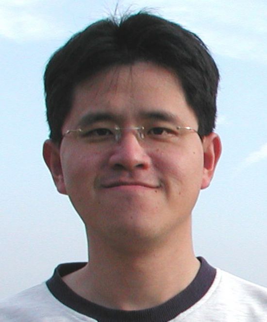
- Ming-Chang Chiang
- MD, PhD
-
Professor, Department of Biomedical Engineering , National Yang Ming Chiao Tung University
E-mail:cgon.chiang@gmail.com
Executive Summary:
Dr. Ming-Chang Chiang received his M.D. degree from National Taiwan University and accomplished his neurology specialist training at National Taiwan University Hospital, Taiwan. He subsequently obtained a Ph.D. degree in Biomedical Engineering from the University of California, Los Angeles, United States, followed by postdoctoral training at the Laboratory of Neuro Imaging at UCLA. Dr. Chiang joined the Department of Biomedical Engineering of National Yang Ming Chiao Tung University, Taiwan, in 2011, and became a professor of Biomedical Engineering in 2022.
Dr. Chiang's research focuses on understanding how chronic neuropathic pain alters the brain by the investigating imaging biomarkers for maladaptive plasticity following chronic neuropathic pain. His work combines advanced neuroimaging techniques such as structural and functional MRI, EEG, and TMS to unravel the complex structural, functional, and electrophysiological changes in the brain connectome resulting from peripheral neuropathic pain.
Research Interests:
Identification of imaging biomarkers for maladaptive plasticity following chronic neuropathic pain.
Applications of structural and functional magnetic imaging (MRI), electroencephalography (EEG), and transcranial magnetic stimulation (TMS) to investigate structural, functional, and electrophysiological alterations of the brain connectome due to the remote effects of neuropathic pain of peripheral origin.
Exploration of brain mechanisms underlying the generation of neuropathic pain and potential treatments of pain by neuromodulation.
Lecture Abstract:
Pain is a common and often the most distressing symptom in many diseases. The assessment of pain usually relies on subjective descriptions and therefore objective biomarkers for pain are needed. To address this problem, neuroimaging techniques, particularly task-based and resting-state functional MRI (fMRI), are applied to provide non-invasive biomarkers for pain. Pain is not mediated simply by a single brain region but involves a complex neural network. Key brain regions involved in pain processing include the anterior cingulate cortex, insular cortex, and primary and secondary somatosensory cortices. Clinically, neuroimaging studies have shown that chronic pain may affect the structure and function of the brain, which is considered a manifestation of maladaptive plasticity. For example, our research found that patients with peripheral neuropathic pain exhibited brain structural alterations such as reductions in brain volume and white matter connectivity of pain-related areas. Moreover, functional connectivity across pain networks in the brain was reduced following chronic neuropathic pain caused by small-fiber neuropathy, and the connectivity reductions predicted the response of anti-neuralgia treatment. Neuroimaging techniques not only provide quantitative information about pain, but also facilitate the assessment of the impact of pain on brain structure and function, and the identification of potential biomarkers to enhance the diagnosis and treatment strategies for patients with chronic pain.
Pain is a common and often the most distressing symptom in many diseases. The assessment of pain usually relies on subjective descriptions and therefore objective biomarkers for pain are needed. To address this problem, neuroimaging techniques, particularly task-based and resting-state functional MRI (fMRI), are applied to provide non-invasive biomarkers for pain. Pain is not mediated simply by a single brain region but involves a complex neural network. Key brain regions involved in pain processing include the anterior cingulate cortex, insular cortex, and primary and secondary somatosensory cortices. Clinically, neuroimaging studies have shown that chronic pain may affect the structure and function of the brain, which is considered a manifestation of maladaptive plasticity. For example, our research found that patients with peripheral neuropathic pain exhibited brain structural alterations such as reductions in brain volume and white matter connectivity of pain-related areas. Moreover, functional connectivity across pain networks in the brain was reduced following chronic neuropathic pain caused by small-fiber neuropathy, and the connectivity reductions predicted the response of anti-neuralgia treatment. Neuroimaging techniques not only provide quantitative information about pain, but also facilitate the assessment of the impact of pain on brain structure and function, and the identification of potential biomarkers to enhance the diagnosis and treatment strategies for patients with chronic pain.
- Time
- Topic
- Speaker
- Moderator
- 14:30-15:00
- Modulation on chronic pain: neurophysiological insights on advancing treatment
- Speaker:
Walter Paulus
- Moderator:
Rou-Shayn Chen
- Walter Paulus
- MD
-
Research Professor, Ludwig Maximilians University Munic
Emeritus Professor of Clinical Neurophysiology, University Medical Center Göttingen
E-mail:wpaulus@gwdg.de
Executive Summary:
WP is Emeritus Professor of Clinical Neurophysiology and former Clinical Director at the Department of Clinical Neurophysiology, University Medical Centre-/ Göttingen, Germany. He started training in neurology in 1978 at the University Hospital for Neurology in Düsseldorf. He spent 6 months at the National Hospital for Neurology and Neurosurgery in London in 1980. After receiving his specialty degree in Neurology he continued at the Alfried Krupp Hospital in Essen and then in 1987 at the Ludwig Maximilian’s University in Munich. He was appointed in Göttingen in 1992. Since his retirement in Göttingen in 2021 he continues research at the Ludwig Maximilians University in Munich, Department of Neurology with EU funded projects. From 2014 to 2018 he was chairman of the European and African Chapter of the International Federation of Clinical Neurophysiology (IFCN), from 2018 to 2022 President of the IFCN, at present being past president. Awards: Price for the best thesis of the University of Düsseldorf (1979); Hans Berger Price of the DGKN (2016); Pierre Gloor Award of the ACNS (2022); European Career Achievement on Non-invasive Brain Stimulation Award of the European Society for Brain Stimulation (2024); International Brain Stimulation Award (IBSA)(2025). Web of Science researcher ID A-3544-2009
WP is Emeritus Professor of Clinical Neurophysiology and former Clinical Director at the Department of Clinical Neurophysiology, University Medical Centre-/ Göttingen, Germany. He started training in neurology in 1978 at the University Hospital for Neurology in Düsseldorf. He spent 6 months at the National Hospital for Neurology and Neurosurgery in London in 1980. After receiving his specialty degree in Neurology he continued at the Alfried Krupp Hospital in Essen and then in 1987 at the Ludwig Maximilian’s University in Munich. He was appointed in Göttingen in 1992. Since his retirement in Göttingen in 2021 he continues research at the Ludwig Maximilians University in Munich, Department of Neurology with EU funded projects. From 2014 to 2018 he was chairman of the European and African Chapter of the International Federation of Clinical Neurophysiology (IFCN), from 2018 to 2022 President of the IFCN, at present being past president. Awards: Price for the best thesis of the University of Düsseldorf (1979); Hans Berger Price of the DGKN (2016); Pierre Gloor Award of the ACNS (2022); European Career Achievement on Non-invasive Brain Stimulation Award of the European Society for Brain Stimulation (2024); International Brain Stimulation Award (IBSA)(2025). Web of Science researcher ID A-3544-2009
Lecture Abstract:
In otherwise pain treatment refractory patients excitatory motor cortex stimulation provides an additional management option. Neurophysiological backgrounds and treatment options as well as protocols will be provided. Some weight will be put on the co-application of drugs such as d-cycloserine. Methodogical aspects of treatment failures will be covered.
In otherwise pain treatment refractory patients excitatory motor cortex stimulation provides an additional management option. Neurophysiological backgrounds and treatment options as well as protocols will be provided. Some weight will be put on the co-application of drugs such as d-cycloserine. Methodogical aspects of treatment failures will be covered.
- Time
- Topic
- Speaker
- Moderator
- 15:00-15:30
- Brain stimulation in neuropathic pain: from central post-stroke pain to peripheral neuropathies
- Speaker:
Koichi Hosomi
- Moderator:
Long-Sun Ro
- Koichi Hosomi
- MD, PhD
-
Specially Appointed Associate Professor, Department of Neurosurgery, The University of Osaka Graduate School of Medicine
E-mail:k-hosomi@nsurg.med.osaka-u.ac.jp
Executive Summary:
Koichi Hosomi is a neurosurgeon specializing in functional neurosurgery and neuromodulation therapies for intractable pain and movement disorders. He graduated from the Faculty of Medicine, The University of Osaka, in 2002 and subsequently completed his training in neurosurgery and earned his Ph.D. In 2010, he was appointed as an assistant professor, and since 2015, he has been an associate professor at the University of Osaka, Japan.
He serves as both a clinician and a researcher at a university hospital and laboratory. He has extensive experience in functional neurosurgery and has conducted numerous experimental and clinical studies on neuromodulation therapies for chronic pain. His research interests include functional neurosurgery, neuromodulation, neuroscience, and central post-stroke pain. He has published multiple papers on clinical trials, neuroimaging studies, and clinical neurophysiology related to both surgical and non-invasive neurostimulation techniques for intractable pain.
Lecture Abstract:
Invasive motor cortex stimulation (MCS) for neuropathic pain was developed by Japanese neurosurgeons in the early 1990s. Initially, it was applied to central post-stroke pain, and later to other chronic pain conditions, including peripheral neuropathic pain. Systematic reviews of MCS have reported a success rate of approximately 50%. In our case series of 39 patients who underwent MCS, 15 patients reported at least a 30% reduction in pain scores during follow-up, with peripheral neuropathic pain tending to respond better than central neuropathic pain. However, MCS is now rarely performed in Japan, as patients prefer non-invasive treatments and it is currently off-label.
Repetitive transcranial magnetic stimulation (rTMS) of the primary motor cortex for intractable pain emerged from clinical experience with MCS. We introduced navigation-guided rTMS in 2003 and have conducted a dozen randomized controlled trials in patients with central and peripheral neuropathic pain, mainly CPSP, to explore optimal target regions, stimulation parameters, and treatment protocol, as well as to verify the safety and efficacy of rTMS therapy for regulatory approval. Although rTMS has not yet been approved as a standard treatment for neuropathic pain in Japan, international therapeutic guidelines and meta-analyses have reported that the high-frequency rTMS of the primary motor cortex is recommended as a third-line treatment for neuropathic pain. In this presentation, I will discuss the current status of MCS and rTMS for neuropathic pain.


